How OsiriX Enhances Veterinary Imaging: A Real-World Use Case
Veterinary medicine increasingly relies on advanced imaging to diagnose, plan treatments, and monitor animal health. Whether it’s evaluating a fracture in a racehorse, assessing a tumor in a cat, or performing pre-surgical planning for a dog, veterinarians need fast, accurate tools that make complex imaging data easy to interpret.
That’s where OsiriX MD comes in — the same high-performance DICOM viewer trusted by hospitals worldwide is also transforming the way veterinary specialists analyze medical images every day.
The Challenge: Complex Cases, Limited Time
A busy veterinary diagnostic center in the United Kingdom handles dozens of CT and MRI studies daily — from small pets to large animals. The diversity of species and anatomy means radiologists need to switch rapidly between protocols, image types, and orientations.
Their biggest challenges were:
-
Slow image rendering and export in large animal CT scans
-
Difficulty sharing 3D reconstructions with surgeons and clients
-
Limited measurement tools in their legacy system
With OsiriX MD, they found an intuitive, high-speed imaging solution that fit perfectly into their existing workflow.
The Solution: Speed, Precision, and Flexibility with OsiriX MD
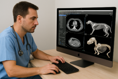
After installing OsiriX MD on macOS workstations, the clinic’s radiologists immediately noticed a difference. The software handled large multi-slice CT scans of equine limbs effortlessly, providing smooth scrolling and high-resolution reconstruction.
Using Volume Rendering and Multiplanar Reconstruction (MPR), the veterinarians could visualize fractures, tumors, and internal structures in precise detail. The ROI tools helped quantify lesion sizes, while 3D models were exported directly for surgical planning or client communication.
One of the most appreciated features? The ability to fuse imaging modalities — for example, combining CT with PET scans in oncology cases to visualize both anatomy and function.
The Impact: Faster Diagnosis, Better Care
The center reported major improvements within weeks of adopting OsiriX MD:
-
50% faster imaging analysis for CT and MRI studies
-
Enhanced collaboration between radiologists and surgeons
-
Improved client communication with high-quality 3D visuals
-
Reduced need for repeat scans, thanks to better image interpretation
Veterinarians particularly praised how intuitive OsiriX is for training interns, enabling faster onboarding and consistent image review standards.
Designed for Veterinary Professionals Too
Though developed for human medicine, OsiriX MD’s robust DICOM engine and visualization tools translate perfectly to the veterinary world. From exotic pets to large animals, OsiriX adapts to any anatomy and imaging modality.
With FDA and CE certifications, it offers the same reliability and quality assurance trusted by medical professionals worldwide — giving veterinarians the confidence to make accurate, data-driven decisions.
Conclusion
OsiriX MD empowers veterinarians to visualize complex anatomy, share insights, and improve diagnostic precision — all in a sleek, high-performance macOS environment. Whether used in small clinics or large referral hospitals, OsiriX is redefining what’s possible in veterinary imaging.
Discover OsiriX MD and see how it can elevate your veterinary practice.