Case Report : Glomus Hypervascularized Tumor
Glomus tumor is a rare benign neoplasm originating from paraganglionic cells of the neural crest developing in the adventitious layer of the vessel. They are non-encapsulated and highly vascularized.
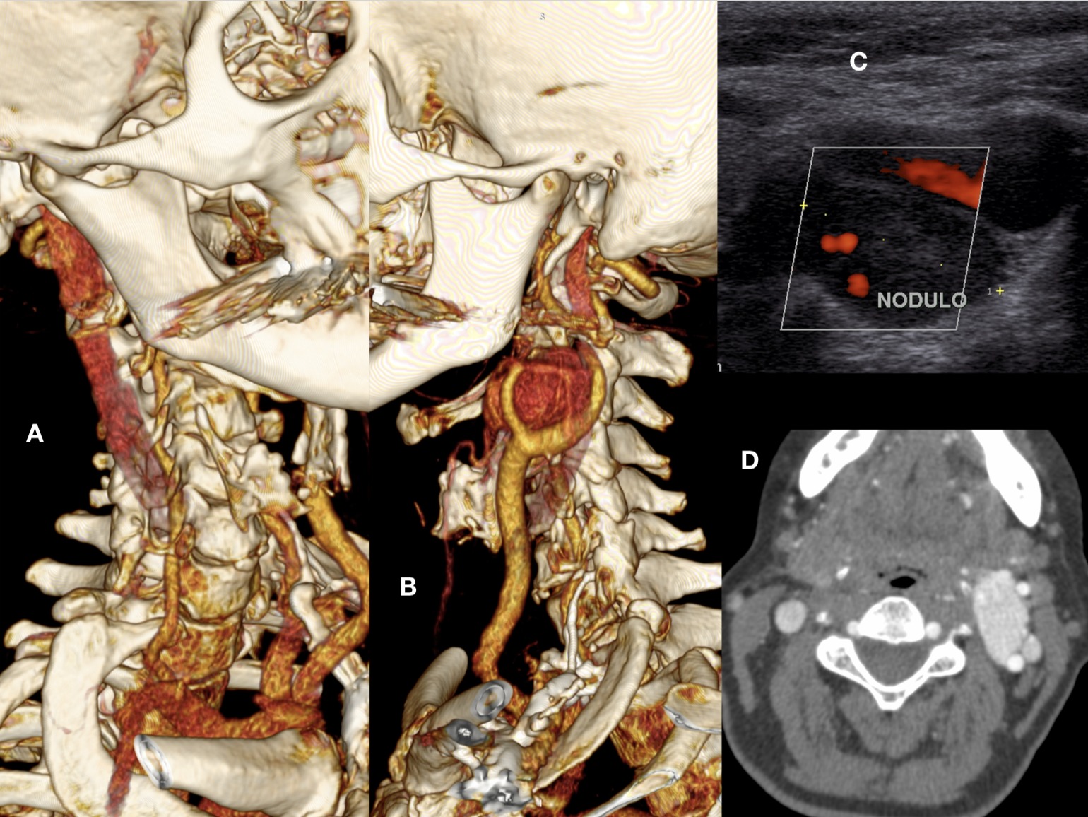
A 64-year-old female patient was identified with a glomus hypervascularized tumor measuring 5 cm posterior to the left carotid bifurcation and contralateral carotid occlusion (Figure 1).
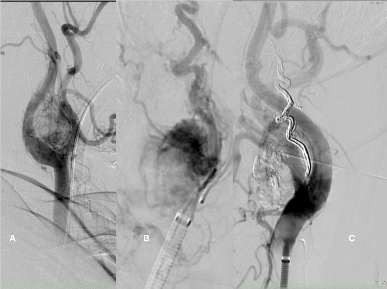
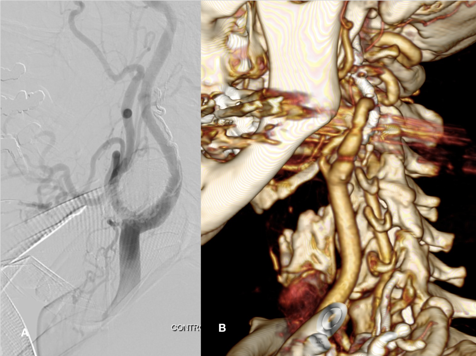
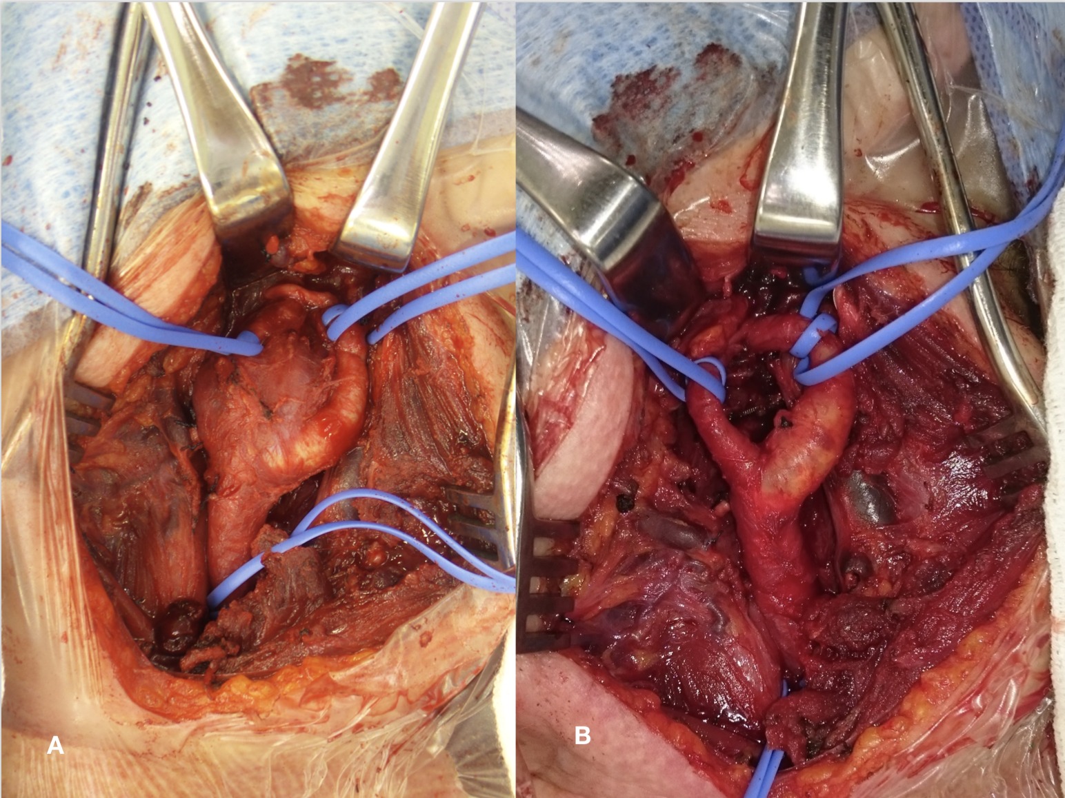
We performed preoperative embolization through endovascular access followed by direct percutaneous puncture, guided by angiography, to fill the remaining area (Figures 2 and 3). After embolization, surgical excision of the tumor with less bleeding was performed and it was easier to find the cleavage plane of the adjacent structures (Figure 4). At late follow-up the patient presents with no tumor recurrence. The tumor was classified as group II, measuring 4 to 6 cm with moderate arterial insertion.
Through this double approach we observed a relative reduction in intraoperative bleeding and improved identification of the cleavage plane, facilitating its excision and avoiding surgical clamping.


