Extend the power of OsiriX with several plugins
These plugins can be downloaded and installed directly from OsiriX: launch OsiriX and open the Plugin Manager in the Plugin menu.
Here is a list of some of these plugins.
 Structured Reporting
Produce complete report in PDF or DICOM PDF, with full diagram & scores.
- Bull’s Eye (for cardiac imaging)
- Prostate (PIRADS)
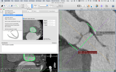
- TAVI Planning (transcatheter aortic valve implantation)
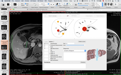
- Liver (Couinaud classification)
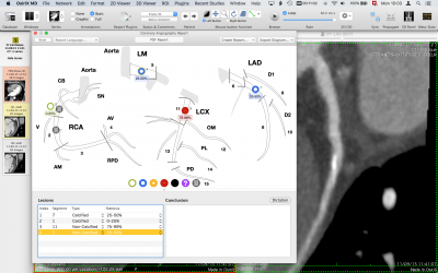
- Coronary Angiography Report
 Bull's Eye
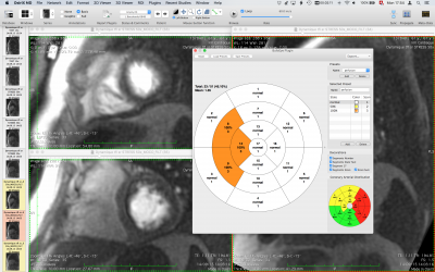
Standardised myocardial segmentation for tomographic imaging of the heart. Nuclear cardiology, echocardiography, cardiovascular magnetic resonance (CMR), cardiac computed tomography (CT), positron emission computed tomography (PET), and coronary angiography are imaging modalities that have been used to measure myocardial perfusion, left ventricular function, and coronary anatomy for clinical management and research. Although there are technical differences between these modalities, all of them image the myocardium and the adjacent cavity. This OsiriX plugin display a short axis representation of the heart, according to the American Heart Association. The diagrams can be exported in PDF, DICOM PDF or PNG images, to be included in the study or added to the report.
 PI-RADS
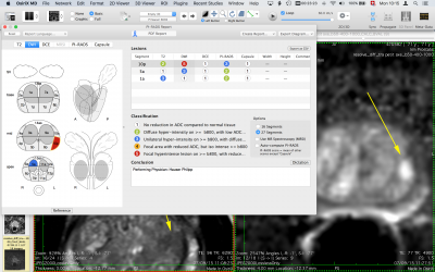
Prostate Imaging-Reporting and Data System (PI-RADS) refers to a structured reporting scheme for prostate cancer. This plugin allows the user to easily represent and compute the PI-RADS score, with a full graphic user interface. The score is assessed on prostate MRI. Images are obtained using a multi-parametric technique including T2 weighted images, a dynamic contrast study (DCE) and DWI. The goal of PI-RADS score is to improve diagnosis and treatment of prostate cancer. The plugin produces a complete report, including diagrams, in high quality DICOM PDF format. This plugin supports PI-RADS 2.0 score. Learn more in the PI-RADS Report Plugin User Manual.
 TAVI Planning
Transcatheter Aortic Valve Implantation Planning
Aortic valve replacement is the mainstay of treatment of symptomatic aortic stenosis. The TAVI procedure requires a comprehensive preinterventional diagnostic workup. Above all, detailed information on the anatomy of the aortic annulus (AA) and the relation of the AA to the coronary arteries is essential to avoid complications. This plugin allows the user to easily, precisely and quickly produces a comprehensive report, including all measurements and screen captures. Learn more in the TAVI Report Plugin User Manual.
 Liver
Liver
Couinaud classification
Lesions (benign or malignant) in the liver require comprehensive description by the radiologist. The liver segmentation facilitates the follow-up and the communication with surgeons, oncologists and gastroenterologists. The oncology CT / MRI report is a vital piece of communication between the radiologist and the treating clinician and often determines patient management. This plugin offers an efficient solution to create RECIST (Response Evaluation Criteria In Solid Tumors) reports The Couinaud classification of liver anatomy divides the liver into eight functionally indepedent segments. Each segment has its own vascular inflow, outflow and biliary drainage. This plugin offers a full graphic user interface to easily create explicit reports and liver diagrams displaying all the lesions. The plugin produces a complete report, including diagrams, in high quality DICOM PDF format. Learn more in the Liver Report Plugin User Manual.
 Coronary Angiography
Easily report the coronaries signifiant and non-signifiant stenosis. It can be used with Coronary CTA studies or Cardiac Catheterisation procedures. Stent icons can also be added to the schema. Learn more in the Coronary Angiography Report Plugin User Manual.
 Ejection Fraction
Compute left ventricule ejection fraction, with mono plane or biplane cine MR sequences.
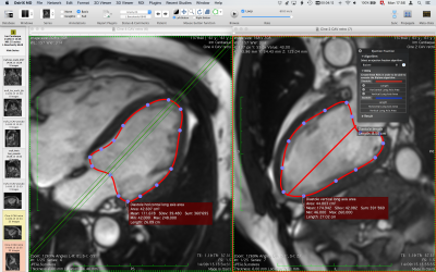
Cardiac MRI allows for measurement of left ventricule ejection fraction, with mono plane or biplane cine MR sequences. This plugin offers a step-by-step semi-automated procedure for the simplified Simpson’s rule. The plugin produces a complete report, including diagrams, in high quality DICOM PDF format.
 Hip Arthroplasty Templates
Pre-operative planning tool for total hip replacement.
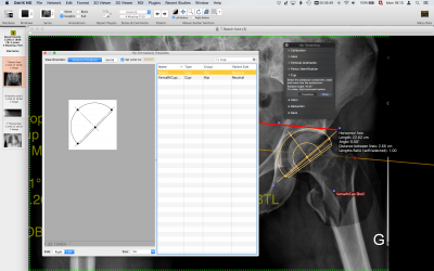
Joint replacement is a well-accepted treatment for arthritic conditions of the hip. Successful surgery requires precise placement of implants such that the function of the joint is optimized biomechanically and biologically. Digital preoperative planning enables the surgeon to select from a library of templates and electronically overlay them on an image. The surgeon can then perform the necessary measurements critical to the templating and preoperative planning process in a digital environment. The preoperative planning process is fast, precise, and cost-efficient, and it provides a permanent, archived record of the templating process
 T2 Mapping
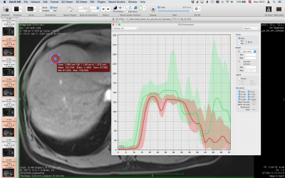
T2 mapping (distribution of T2 values) is a notable MRI technique for evaluating water contents in tissues. This plugins produces T2 values from multi echo T2 series. T2 mapping might be a sensitive method for the detection of early cartilage degeneration. T2 mapping is also used for detection of active myocarditis.
 Volume calculator
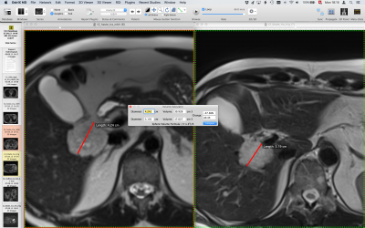
Easily estimate a 3D volume from a diameter.
This plugin offers an efficient interface to compute the volume from a diameter. It can also compares 2 volumes to get the percentage change. The percentage change in volume is a more accurate measurement to treatment response, compared to lesions diameter
![]()
Third Party Plugins
The following plugins are not distributed with OsiriX, but can be obtained through their respective manufacturers. Additional fees may apply.
Measure New Predictive MRI Imaging Biomarkers Of Cardiovascular Events
ArtFun+ is an MDR CE class 2a MRI analysis plugin designed to measure arterial stiffness and flow biomarkers, such as Ascending Aortic Distensibility and Aortic Pulse Wave Velocity. These biomarkers are commonly recognized as major risk factors for cardiovascular diseases and significant predictors of cardiovascular mortality.
In combination with other risk factors, these advanced cardiovascular imaging biomarkers improve the understanding of a patient’s cardiovascular risk and help quantify cardiovascular aging. They complement routine MRI exams by enabling a personalized prognosis of cardiovascular health.
ArtFun+ is the most clinically validated and widely published technology (with over 70 publications) for measuring arterial stiffness biomarkers by MRI (validated in the Multi-Ethnic Study of Atherosclerosis clinical trial).
Top Features
- Segmentation of the great arteries, such as the aorta
- Extraction of arterial flow and area curves
- Extraction of arterial centerline
- Measurement of Transit Time between flow curves
- Measurement of arterial stiffness biomarkers (PWV, distensibility) and flow indices
- Export the results in CSV format
 IB Neuro
Standardized quantitative MR DSC perfusion maps
Exclusive to IB Neuro, offers the most accurate and repeatable MR DSC perfusion approach available. This plugin automatically generates common perfusion parameters such as cerebral blood flow (CBF), mean transit time (MTT), and relative cerebral blood volume (rCBV). For patients with brain tumors, rCBV is becoming an essential imaging biomarker for assisting physicians in identifying tumor grade (aggressiveness), distinguishing progression from pseudo-progression and tumor from treatment effect, as well as monitoring treatment response. Given the proven accuracy and repeatability, IB Neuro also fosters the assessment of new treatment therapies and it is a core technology required for generating Fractional Tumor Burden (FTB) maps.
 IB DCE
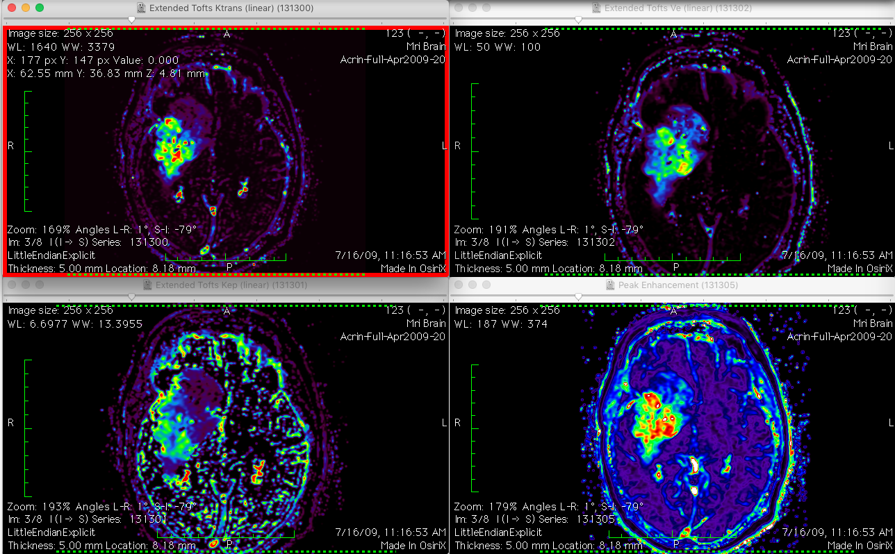
Easily Compute MR DCE parameters
Accurate implementation of the Tofts, Extended Tofts, and Patlak models, IB DCE offers an intuitive solution for generating relevant perfusion and permeability parameters (Ktrans, Ve, Vp, T10, Initial Slope, Time to Peak, etc.). This whole-body application consists of the same user interface as other OsiriX plugins made by Imaging Biometrics.
 IB Diffusion
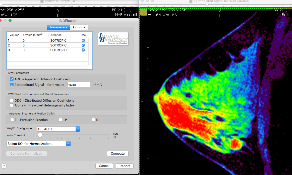
Generate ADC maps from DWI datasets
Diffusion-weighted imaging (DWI) is a powerful MRI method which probes abnormalities of tissue structure by detecting microscopic changes in water mobility at a cellular level beyond what is available with other imaging methods. Accordingly, DWI has the potential to identify pathology before gross anatomic changes are evident on standard anatomical brain images. This OsiriX plugin generates apparent diffusion coefficient (ADC) maps, and other diffusion parameters, from routinely acquired DWI datasets.
 IB Delta Suite
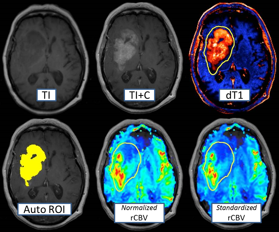
Standardize, Register, Subtract, Quantify, and Visualize
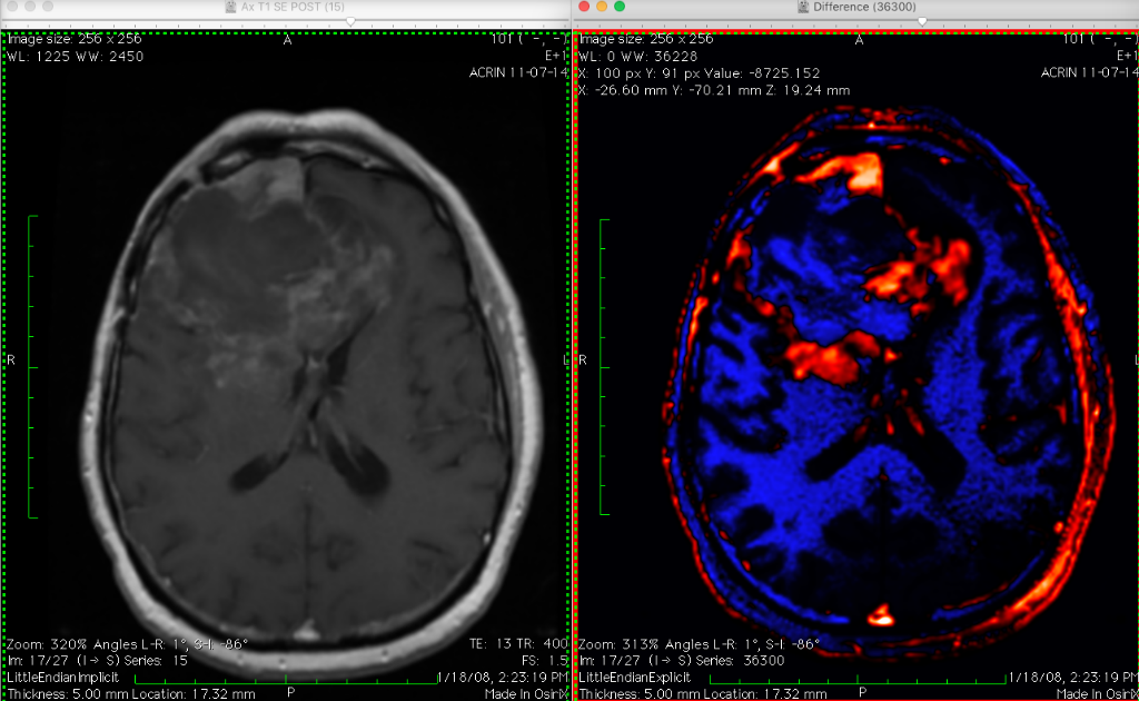
The ability to accurately determine regions of contrast-enhancing (T1w + contrast agent) tumor volume on MR datasets is problematic due to inherent variabilities and inconsistencies. For example, non contrast-enhancing tissues, such as post-surgical blood products, can also appear bright on post contrast images. Even with expert readers, the inter-reader agreement is usually no better than 50-60%. This OsiriX plugin offers the ability to generate “delta T1” (dT1) maps. These maps use exclusively licensed technology to generate quantitatively consistent images between time points, scanner platforms and field strengths, and patients. In general, IB Delta Suite performs image registration, ROI thresholding, class map generation, image exporting, and more.
 Dental3DPlugin
Your planning tool for dental implants treatments.
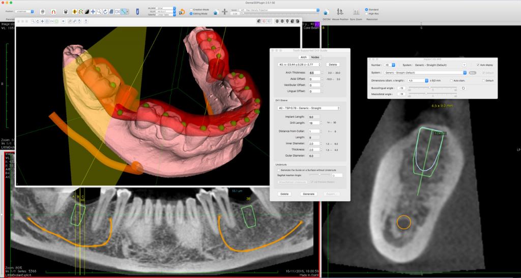
Dental3DPlugin extends both the OsiriX 3D Curved MPR Viewer and the 3D Surface Rendering Viewer. It allows the user to plan dental implant treatments and design surgical drill guides. The guides designed with the plugin can easily be exported as STL files and printed out on the 3D printer of your choice.
Freely available in OsiriX’s Plugin Manager. Launch OsiriX, open the Plugins Menu and choose Plugins Manager.
OsiriX Plugin Development Guide
Developers wanting to create their own plugins, should start by reading our OsiriX Plugin Development Guide.


 ROI Enhancement